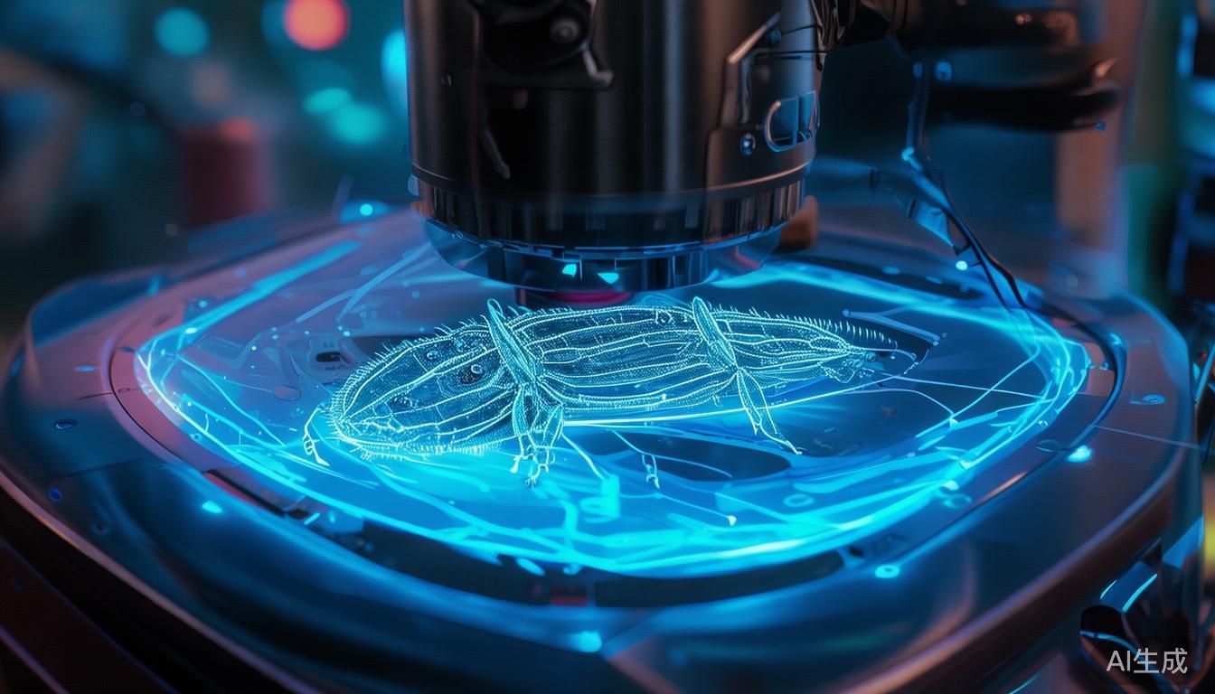
撕开微观层面:飞秒激光横切昆虫
显微镜领域长期以来一直是静态观察的领域,科学家们通过肉眼无法察觉的尺度,窥探生命体的复杂结构。从叶片的精细叶脉到细胞内的复杂机械装置,传统方法依赖染色、固定和切片来揭示隐藏的细节。但如果有一种方法不仅能观察,还能精确地雕刻昆虫外骨骼的每一层纤维,同时又不破坏其内部精细结构呢?这正是飞秒激光横切昆虫这一突破性技术登场的地方,它为我们展示了未来微观操控将像活检一样成为常规操作的前景。
飞秒激光的力量
要理解这项技术的意义,首先必须了解什么是飞秒激光。与发射连续波的传统激光不同,飞秒激光产生极其短暂、高强度的光脉冲——仅持续一千万亿分之一秒(10^-15秒)。这种短暂的时间允许激光以高度可控的方式与材料相互作用,最大限度地减少热损伤,并实现微尺度上的精确、非侵入性切割。
飞秒激光的精妙之处在于它们能够产生光化学效应而非热效应。当激光脉冲与材料相互作用时,会产生一波自由电子,这些电子迅速膨胀,形成一种称为"光化学爆炸"的微观爆炸。这种爆炸有效地在外材料中烧出一个洞,而不会加热周围区域,使其成为处理精细生物样本的理想工具。
昆虫外骨骼的挑战
昆虫为微观操控带来了独特的挑战。它们的由几丁质组成的外骨骼非常坚硬且不透水,旨在抵御环境压力和捕食者。传统方法在制备昆虫样本进行显微镜观察时,通常涉及强化学物质或机械研磨,这可能会损坏或扭曲昆虫内部的精细结构。
对于研究昆虫解剖学的科学家来说,这是一个重大障碍。理解外部结构与内部器官之间的关系需要高分辨率成像,但制备过程本身可能会损害样本的完整性。这就是飞秒激光提供革命性解决方案的地方。
技术操作过程
使用飞秒激光横切昆虫的过程涉及几个关键步骤:
-
样本固定:首先将昆虫固定,并将其放置在稳定的基板上,以防止在激光操作过程中移动。
-
激光编程:通过先进软件,研究人员可以编程激光沿特定平面进行精确切割。这允许创建横截面,揭示内部解剖结构,而无需机械力。
-
脉冲控制:激光设置为发送一系列经过精心校准的脉冲,每个脉冲都会产生一个微观爆炸,精确地切割外骨骼并进入内部结构。
-
成像:完成横切后,可以使用电子显微镜立即对样本进行成像,提供高分辨率的内部解剖结构视图,而不会因传统制备方法而产生变形。
来自Hacker News的链接使用飞秒激光在电子显微镜下横切昆虫展示了这一过程的迷人之处。观看激光如何精确地切割昆虫的外骨骼,揭示其下肌肉、神经和器官的复杂网络。视频中展示的精确性和控制力突出了这项技术在未来推动昆虫生物学研究进展的潜力。
相比传统方法的优点
飞秒激光方法相比传统的昆虫制备方法具有以下几个优点:
-
损伤最小:通过避免热效应,激光保留了昆虫内部的精细结构,提供了更准确和详细的观察结果。
-
速度和精度:激光的高速特性允许快速横切,而精度确保研究人员可以针对特定感兴趣区域。
-
减少化学使用:与传统方法依赖强化学物质进行组织透明化和染色不同,激光技术最大限度地减少了化学处理的需求,使其更加环保。
-
提高成像质量:由于没有机械应力,内部结构保持其自然状态,从而获得更高质量的成像结果。
科学和工业中的应用
这项技术的潜在应用非常广泛,涵盖多个领域:
生物学和昆虫学
对于生物学家和昆虫学家来说,能够非侵入性地解剖昆虫为研究开辟了新的途径。研究昆虫器官的结构-功能关系、理解外骨骼的发育以及调查运动的生物力学现在可以进行前所未有的细节。这可能导致在害虫控制等领域的突破,其中理解昆虫解剖学可以带来更有效和更有针对性的干预措施。
医学研究
飞秒激光横切技术的原理也可以应用于医学研究。例如,研究携带疟疾或登革热等疾病的昆虫的解剖结构,可以提供关于媒介生物学的见解,并帮助制定新的疾病预防策略。此外,该技术还可以适应用于人类组织分析,为传统活检方法提供非侵入性替代方案。
材料科学
飞秒激光的精度使其成为材料科学研究的有价值工具。通过创建昆虫外骨骼的精确横截面,研究人员可以研究几丁质和其他天然聚合物的材料特性,有可能开发出具有工程和制造应用的新仿生材料。
挑战和未来方向
尽管潜力巨大,但用于横切昆虫的飞秒激光技术仍处于早期阶段。为了使其成为研究实验室的主流工具,需要解决几个挑战:
-
成本和可及性:高性能飞秒激光很昂贵,这可能会限制其在较小研究机构或发展中国家的可用性。
-
样本大小和多样性:该技术主要在小昆虫上进行了测试。将其扩展到更大的样本或应用于更广泛的昆虫物种需要进一步改进。
-
长期影响:虽然激光对样本的即时影响很小,但了解对组织完整性的长期影响对于确保结果的可靠性至关重要。
-
与其他技术的整合:将激光技术与荧光显微镜或3D重建等其他成像方式相结合,可以提供更全面的昆虫解剖学见解。
未来的研究可能会集中于解决这些挑战并扩展该技术的功能。随着技术的成熟,我们可以期待其在学术和工业环境中的更广泛采用,推动昆虫生物学及其他领域的发现。
总结
使用飞秒激光横切昆虫代表了对微观操控和成像的重大飞跃。通过提供一种非侵入性、精确且损伤最小的昆虫解剖学研究方法,这项技术为多个学科的研究开辟了新的可能性。从增进我们对昆虫生物学的理解到有可能彻底改变医学成像,应用范围广泛且令人兴奋。随着我们继续推动微观尺度可能性的边界,飞秒激光作为创新之光,以前所未有的清晰度照亮了昆虫世界的隐藏奇迹。
Peeling Back the Microscopic Layers: Cross-Sectioning Insects with a Femtosecond Laser
The world of microscopy has long been a realm of static observation, where scientists peer into the intricate structures of life at scales invisible to the naked eye. From the delicate veins of a leaf to the complex machinery within a cell, traditional methods have relied on staining, fixing, and slicing to reveal hidden details. But what if there was a way to not just observe, but to precisely carve into the very fabric of an insect’s exoskeleton without destroying its delicate internal structures? This is where the groundbreaking technique of cross-sectioning insects with a femtosecond laser steps in, offering a glimpse into a future where microscopic manipulation becomes as routine as a biopsy.
The Power of the Femtosecond Laser
To understand the significance of this technique, it’s essential to first grasp what a femtosecond laser is. Unlike conventional lasers that emit light in continuous waves, femtosecond lasers produce incredibly short, high-intensity bursts of light—on the order of one quadrillionth of a second (10^-15 seconds). This brief duration allows the laser to interact with materials in a highly controlled manner, minimizing thermal damage and enabling precise, non-invasive cutting at the microscale.
The beauty of femtosecond lasers lies in their ability to create a photochemical effect rather than a thermal one. When the laser pulse interacts with a material, it generates a wave of free electrons that rapidly expand, creating a microscopic explosion known as a "photochemical explosion." This explosion effectively "burns" a hole in the material without heating the surrounding area, making it an ideal tool for delicate biological samples.
The Challenge of Insect Exoskeletons
Insects present a unique challenge for microscopic manipulation. Their exoskeletons, composed primarily of chitin, are incredibly tough and impermeable, designed to protect against environmental stressors and predators. Traditional methods of preparing insect samples for microscopy often involve harsh chemicals or mechanical grinding, which can damage or distort the delicate internal structures.
For researchers studying insect anatomy, this poses a significant hurdle. Understanding the relationship between external structures and internal organs requires high-resolution imaging, but the preparation process itself can compromise the integrity of the sample. This is where the femtosecond laser offers a revolutionary solution.
The Technique in Action
The process of cross-sectioning an insect with a femtosecond laser involves several key steps:
-
Sample Stabilization: The insect is first immobilized and placed on a stable substrate to prevent movement during the laser procedure.
-
Laser Programming: Using advanced software, researchers can program the laser to create precise cuts along specific planes. This allows for the creation of cross-sections that reveal the internal anatomy without the need for mechanical force.
-
Pulse Control: The laser is set to deliver a series of carefully calibrated pulses, each one creating a microscopic explosion that carves a precise path through the exoskeleton and into the internal structures.
-
Imaging: Once the cross-section is complete, the sample can be immediately imaged using an electron microscope, providing high-resolution views of the internal anatomy without the distortion caused by traditional preparation methods.
The video linked from Hacker News Cross sectioning insects in an electron microscope with a femtosecond laser offers a fascinating glimpse into this process. Watch as the laser meticulously carves through the insect’s exoskeleton, revealing the intricate network of muscles, nerves, and organs beneath. The precision and control demonstrated in the video highlight the potential of this technique for advancing our understanding of insect biology.
Advantages Over Traditional Methods
The femtosecond laser approach offers several advantages over conventional methods of insect preparation:
-
Minimal Damage: By avoiding thermal effects, the laser preserves the delicate structures within the insect, providing more accurate and detailed observations.
-
Speed and Precision: The high-speed nature of the laser allows for rapid cross-sectioning, while the precision ensures that researchers can target specific areas of interest.
-
Reduced Chemical Use: Unlike traditional methods that rely on harsh chemicals for tissue clearing and staining, the laser technique minimizes the need for chemical processing, making it more environmentally friendly.
-
Improved Imaging Quality: The lack of mechanical stress means that the internal structures remain in their natural state, leading to higher-quality imaging results.
Applications in Science and Industry
The potential applications of this technique are vast and span across multiple fields:
Biology and Entomology
For biologists and entomologists, the ability to non-invasively dissect insects opens up new avenues for research. Studying the structure-function relationships in insect organs, understanding the development of exoskeletons, and investigating the biomechanics of movement can now be done with unprecedented detail. This could lead to breakthroughs in areas such as pest control, where understanding insect anatomy could lead to more effective and targeted interventions.
Medical Research
The principles of femtosecond laser cross-sectioning can also be applied to medical research. For instance, studying the anatomy of insects that carry diseases like malaria or dengue fever can provide insights into vector biology and help develop new strategies for disease prevention. Additionally, the technique could be adapted for use in human tissue analysis, offering a non-invasive alternative to traditional biopsy methods.
Materials Science
The precision of the femtosecond laser makes it an invaluable tool for materials science research. By creating precise cross-sections of insect exoskeletons, researchers can study the material properties of chitin and other natural polymers, potentially leading to the development of new biomimetic materials with applications in engineering and manufacturing.
Challenges and Future Directions
Despite its promising potential, the femtosecond laser technique for cross-sectioning insects is still in its early stages. Several challenges need to be addressed to make it a mainstream tool in research laboratories:
-
Cost and Accessibility: High-powered femtosecond lasers are expensive, which may limit their availability in smaller research institutions or developing countries.
-
Sample Size and Diversity: The technique has primarily been tested on small insects. Scaling it up to larger specimens or applying it to a wider variety of insect species will require further refinement.
-
Long-Term Effects: While the immediate impact of the laser on the sample is minimal, understanding the long-term effects on the integrity of the tissue is crucial for ensuring the reliability of the results.
-
Integration with Other Techniques: Combining the laser technique with other imaging modalities, such as fluorescence microscopy or 3D reconstruction, could provide even more comprehensive insights into insect anatomy.
Future research will likely focus on addressing these challenges and expanding the capabilities of the technique. As the technology matures, we can expect to see more widespread adoption in academic and industrial settings, driving new discoveries in insect biology and beyond.
Takeaway
The ability to cross-section insects with a femtosecond laser represents a significant leap forward in microscopic manipulation and imaging. By offering a non-invasive, precise, and minimally damaging method for studying insect anatomy, this technique opens up new possibilities for research across multiple disciplines. From advancing our understanding of insect biology to potentially revolutionizing medical imaging, the applications are vast and exciting. As we continue to push the boundaries of what’s possible at the microscale, the femtosecond laser stands as a beacon of innovation, illuminating the hidden wonders of the insect world with unprecedented clarity.